Apply Now Programmes Virtual Tour CT-SET 2026 Ph.D
Admissions
Open 2026-27
Apply now
Apply Now
Home  /  Programmes  /  Bachelor in Medical Radiology and Imaging Technology (Lateral Entry)

Bachelor in Medical Radiology and Imaging Technology (Lateral Entry)

Duration: 3 Years Category: Graduate Programs

Programme Details

About the Programme

B.Sc. in Medical Radiology & Imaging Technology is a 3-year undergraduate course. It provides rigorous foundations of core Medical Radiology and Imaging Technology, combination of theoretical core concepts with practical training of radiological skills which equips the students to be industry ready. Objective of this programme is to compete with well-established academic institutions in India and abroad.

Industry Immersion

Those who choose this stream are going to study about Medical Radio Imaging Technology, Equipment’s, Emergency Care in Radiology etc. Those who get their placement will be known as Radiology Assistants/Technicians & will Assist an Radiologist in diagnosing various diseases with the help of imaging the body parts by various machines. This Program will create a great source of manpower that can aid in our health sector especially in Radiology, Emergency & Operation Theatres. Radiology Technologists will provide safe & quality patient care by using their technical & critical thinking while Examining the patient. This Program will give students knowledge of basic Science like Anatomy, Physiology, Microbiology& Pathology etc.

Eligibility Criteria

Candidates passed minimum 2 year full time diploma in RIT/MIT program from the Government Boards and recognized by State/Central University, with 10+2 with Physics, Chemistry & Biology with 50% Marks

Register Now

Why Choose CT University

Why CT University is the Right Choice for Your Future

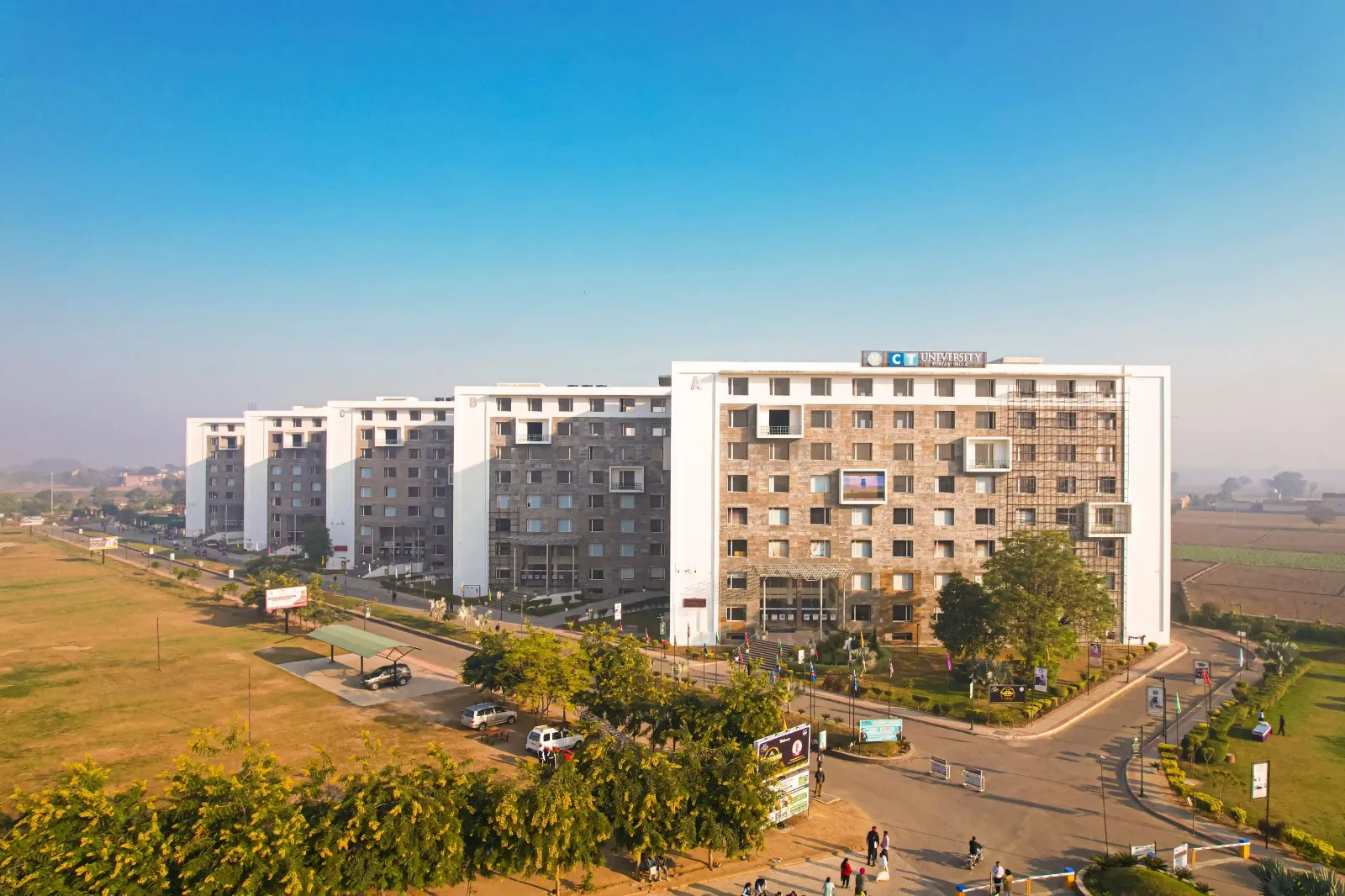
CT University Campus

At CT University, we combine industry-integrated learning and global academic partnerships, backed by the CT Group's 30+ year legacy, to prepare students for careers that actually last.

Industry-oriented
learning

Modern campus
facilities

Academic excellence &
mentorship

Global opportunities &
placements

Fee Structure & Scholarships

Program Fee Details

Fee Type Amount
Programme Fees (per Semester) ₹60000
Examination Fees ₹3000

Merit Based Scholarship Scheme

Unlock up to 90% Scholarship through CTSET, with scholarships worth Rs 40 Crore! Get access to top-quality education at CT University, Ludhiana — without the financial burden.

Slab >=60% - 74.99% >=75% - 89.99% >=90% & Above
Fee ₹55000 ₹50000 ₹45000

Students can avail these slots depending on the marks they have scored. Each slot reflects a different academic range, helping students understand where they stand and what benefits they qualify for.

Course Curriculum

1ST SEMESTER SUBJECTS

Provides comprehensive knowledge of the structure and organization of the human body with emphasis on anatomical systems relevant to medical imaging and radiologic practice.
Course Outcome:
CO1: Describe the general anatomy of human body
CO2: Explain normal disposition of various structures and organs in the body and its clinical correlation
CO3: Describe the microscopic structure of various tissues
CO4: Determine the topography of various structures on the surface of the body
CO5: Identify and locate structures of the body
CO6: Identify organs and tissues under microscope
CO7: Point out various features of appearance of normal body in skiagrams

Introduces the normal functions and physiological processes of major human body systems essential for understanding clinical conditions and medical imaging.
Course Outcome:
CO1: Explain the normal functioning of organs and systems.
CO2: Understand the interrelationships and interactions among various organs and systems for maintaining homeostasis.
CO3: Assess the relative contribution of each organ systems toward the maintenance of constant internal environment
CO4: Differentiate between normal and abnormal functioning of organs and systems.
CO5: Understand physiological basis of pathogenesis and treatment of diseases and disorders.
CO6: Apply the physiological basis in the field of allied health care

Introduces the fundamental principles of radiation physics, properties of radiation, radiation production, interaction with matter, and basic radiation safety concepts.
Course Outcome:
CO1: Describe general physics related to imaging
CO2: Differentiate between within general radiation
CO3: Identify construction of radiology equipment’s
CO4: Interpret quality of control of radiology equipment’s
CO5: Differentiate between x-ray equipment’s and other radiology related equipment’s
CO6: Describe production of x-rays
CO7: Describe circuit system of radiology equipment’s

Provides an overview of healthcare systems, healthcare professionals, patient care, infection control, safety, and the role of medical imaging professionals in healthcare delivery.
Course Outcome:
CO1: Explain the fundamental concepts, structure, and functions of the healthcare system.
CO2: Describe the roles and responsibilities of healthcare professionals within multidisciplinary healthcare teams.
CO3: Explain basic concepts of patient care, health promotion, disease prevention, and healthcare delivery.
CO4: Describe principles of infection prevention, patient safety, and standard precautions in healthcare settings.
CO5: Explain the importance of communication, documentation, confidentiality, and patient-centered care.
CO6: Apply basic healthcare principles to provide safe, ethical, respectful, and patient-centered radiological services.

Develops knowledge of medical terminology, clinical documentation, patient records, and systematic record-keeping practices used in healthcare and radiology services.
Course Outcome:
CO1: Explain the basic principles and structure of medical terminology used in healthcare and radiology.
CO2: Identify and interpret common medical terms, abbreviations, prefixes, suffixes, and root words.
CO3: Describe the terminology associated with major anatomical systems, diseases, diagnostic procedures, and radiological investigations.
CO4: Demonstrate accurate preparation, maintenance, and organization of patient and imaging records.
CO5: Explain the importance of confidentiality, accuracy, completeness, and security of healthcare records.
CO6: Apply appropriate medical terminology and record-keeping practices in routine radiological and healthcare settings.

Develops basic computer literacy and information science skills for digital documentation, data management, communication, and information handling in healthcare and medical imaging.
Course Outcome:
CO1: Explain the fundamental concepts, components, and applications of computers in healthcare.
CO2: Demonstrate basic skills in operating computer systems, software applications, and digital healthcare tools.
CO3: Describe the principles of data management, information storage, retrieval, and security in healthcare environments.
CO4: Explain the basic concepts and applications of hospital information systems and radiology information systems.
CO5: Describe the role of digital imaging, electronic health records, and information technology in modern healthcare.
CO6: Apply basic computer and information-management skills for accurate, secure, and efficient radiological documentation and communication.

Introduces medical law, professional ethics, patient rights, confidentiality, informed consent, legal responsibilities, and ethical decision-making in healthcare practice.
Course Outcome:
CO1: Explain the fundamental concepts, principles, and significance of medical law and ethics in healthcare.
CO2: Describe the legal responsibilities, professional duties, and scope of practice of radiology and imaging professionals.
CO3: Explain the principles of patient rights, informed consent, confidentiality, privacy, and professional accountability.
CO4: Describe ethical principles related to patient care, radiation exposure, diagnostic imaging, and professional practice.
CO5: Explain the importance of documentation, negligence prevention, professional conduct, and compliance with applicable healthcare regulations.
CO6: Apply legal and ethical principles to provide safe, respectful, confidential, and responsible radiological services.

Develops professional behaviour, values, communication, teamwork, accountability, empathy, and ethical conduct required for competent and patient-centred radiology practice.
Course Outcome:
CO1: Explain the fundamental principles of professionalism and professional values in healthcare practice.
CO2: Describe the importance of integrity, accountability, empathy, respect, and responsibility in patient care.
CO3: Demonstrate effective communication and interpersonal skills while interacting with patients and healthcare professionals.
CO4: Explain the importance of teamwork, collaboration, leadership, and professional behaviour in healthcare settings.
CO5: Demonstrate ethical conduct, confidentiality, respect for diversity, and patient-centered behaviour.
CO6: Apply professional values and ethical behaviour to provide safe, compassionate, and effective radiological services.

Introduces fundamental principles of management, organization, leadership, resource utilization, teamwork, and administrative practices relevant to healthcare and medical imaging departments.
Course Outcome:
CO1: Explain the fundamental concepts, principles, and functions of management in healthcare organizations.
CO2: Describe the principles of planning, organizing, staffing, directing, coordinating, and controlling healthcare activities.
CO3: Explain the importance of leadership, communication, motivation, and teamwork in healthcare management.
CO4: Describe basic principles of resource management, inventory control, scheduling, and workflow management in radiology departments.
CO5: Explain the importance of quality management, patient safety, risk management, and continuous improvement in healthcare services.
CO6: Apply basic management principles to support efficient, safe, and effective functioning of radiological services.

Develops English language, communication, interpersonal, presentation, and professional communication skills required for effective interaction with patients and healthcare professionals.
Course Outcome:
CO1: Demonstrate basic proficiency in listening, speaking, reading, and writing for professional communication.
CO2: Develop appropriate vocabulary, grammar, and language skills for academic and healthcare communication.
CO3: Demonstrate effective verbal and non-verbal communication with patients, colleagues, and healthcare professionals.
CO4: Apply appropriate communication skills for taking basic patient information and providing procedural instructions.
CO5: Demonstrate effective written communication through reports, records, emails, and professional documentation.
CO6: Apply communication skills to promote patient understanding, teamwork, professionalism, and effective healthcare delivery.

Provides supervised clinical exposure to radiology services, patient care, professional conduct, basic imaging procedures, radiation safety, departmental workflow, and healthcare team practices.
Course Outcome:
CO1: Demonstrate professional behaviour, discipline, and appropriate communication within the radiology department.
CO2: Identify the basic workflow, organization, equipment, and functions of different sections of a radiology department.
CO3: Demonstrate appropriate patient identification, preparation, positioning, and communication under supervision.
CO4: Apply basic principles of radiation protection, infection prevention, patient safety, and standard precautions during clinical exposure.
CO5: Observe and assist with routine radiological procedures under the supervision of qualified radiology professionals.
CO6: Maintain appropriate clinical records and demonstrate confidentiality, teamwork, ethical conduct, and professional responsibility during studentship.

2ND SEMESTER SUBJECTS

Introduces basic concepts of microorganisms, infection, transmission, sterilization, disinfection, and infection control practices relevant to radiology and healthcare settings.
Course Outcome:
CO1: Describe the structure, classification, morphology and growth of bacteria
CO2: Describe the methods sterilization and disinfection and its applications
CO3: Explain the concepts of immunity, hypersensitivity and immunization
CO4: Describe Nosocomial infections and methods for prevention of Hospital acquired infections
CO5: Describe the management of biomedical waste
CO6: List the common fungi and viruses and explain their importance

Introduces fundamental concepts of carbohydrates, proteins, lipids, enzymes, vitamins, minerals, metabolism, and their relevance to human health and clinical practice.
Course Outcome:
CO1: Understand the responsibility of health care personals and hazardsfaced in the clinical laboratory
CO2: Explain the different types, use, care and maintenance of laboratory apparatus and instruments.
CO3: Understand the fundamental chemistry and knowledge of different solutions
CO4: Understand what acids, bases, salts and indicators are and also know about acid base balance
CO5: Describe the sample collection procedure to analyse various biochemical parameters
CO6: Describe assimilation of nutrients and consequences of malnutrition
CO7: Understand the different functional tests like LFT (Liver function test), RFT (Renal function test)
CO8: Understand the overview of tumor markers, cardiac markers, blood sugar and GTT, lipid profile and diagnostic enzymology
CO9: Describe the applications of radioisotopes

Covers the principles of conventional radiography, X-ray production, radiographic equipment, image formation, accessories, quality control, and radiation safety.
Course Outcome:
CO001: Describe the structure and working of x-ray tube, production of x-rays
CO002: Describe the types of x-ray tube and heat dissipation methods
CO003: Explain the x-ray generator circuits
CO004: Describe the different circuit types
CO005: Describe the meters and exposure timers
CO006: List the control of scattered radiation
CO007: Describes about the fluoroscopy
CO008: Explains about the care and maintenance of x-ray equipment’s

Develops foundational skills in patient positioning, radiographic positioning principles, anatomical landmarks, image evaluation, and safe acquisition of routine radiographic examinations.
Course Outcome:
CO1: Understand the basic patient positioning during radiographic investigation.
CO2: Apply special positioning skillsfor different pathological and physical conditions.
CO3: Application of equipments while working in radiology departments.
CO4: Choose proper position during radiography.
CO5: Explain relative positions of x-ray tube and patient relevant exposure factors during radiography.
CO6: Explain the use of accessories.
CO7: Explain the anatomic and physiological basis of the procedure to be undertaken.
CO8: Explain the radiographic appearances of both normal and common abnormal conditions.

Provides supervised clinical experience in radiology departments, developing competencies in patient care, radiographic procedures, equipment handling, radiation protection, professional conduct, and departmental workflow.
Course Outcome:
CO1: Demonstrate professional behaviour, communication, discipline, and teamwork within the clinical radiology environment.
CO2: Demonstrate appropriate patient identification, preparation, positioning, and communication under supervision during routine radiographic procedures.
CO3: Assist in the operation of conventional radiography equipment and follow established departmental protocols under supervision.
CO4: Apply radiation protection, infection prevention, patient safety, and standard precautions during clinical radiography procedures.
CO5: Observe and assist with routine radiographic examinations and evaluate basic image quality under the supervision of qualified professionals.
CO6: Maintain appropriate clinical records and demonstrate confidentiality, ethical conduct, teamwork, and professional responsibility throughout the clinical studentship.

3RD SEMESTER SUBJECTS

Introduces the fundamental concepts of disease processes, cellular changes, inflammation, infection, and common pathological conditions relevant to medical imaging and patient care.
Course Outcome:
CO1: Define the term “Disease” or concepts of Diseases.
CO2: Define, classify diseases and the medical terms used.
CO3: Describe the cause and mechanism of a few common diseases they come across during their routine work.
CO4: Common changes seen in these diseased persons in different organs/tissues/bodyfluids.
CO5: Names of the common laboratory tests done to diagnose the diseases like examination of urine, blood, other body fluids and tissues.
CO6: Enumerate the proper methods of collection, preservation and delivery of the samples to the respective laboratories.
CO7: Describe the procedures of procuring the whole blood or blood components from blood bank and the complications of blood transfusion.

Develops advanced skills in radiographic positioning, anatomical landmarks, patient preparation, image evaluation, and safe performance of routine and specialized radiographic examinations.
Course Outcome:
CO1: Prepare management and positioning of patients
CO2: Correlate of indications, contraindications of the patient
CO3: Understand the patient preparations needed before any radiological examination.
CO4: Generalize knowledge of post procedural care.
CO5: Students will be able position the patients for radiological procedures.
CO6: Knowledge of image quality in radiological images.
CO7: Management of patientsin radiology department for various procedures.
CO8: Ability to handle emergency situations in radiology department.

Covers radiographic image formation, processing techniques, image quality, digital radiography, quality assurance, artifacts, and methods for optimizing diagnostic image quality.
Course Outcome:
CO1: Know basic physics of radiography processing system
CO2: Describe construction and working of film, intensifying screen, cassette, dark room and automatic processor
CO3: Explain radiographic film Processing chemistry
CO4: Discuss the factors affecting image quality in radiographic image and their application
CO5: Operate the workflow in x-ray imaging
CO6: Apply knowledge for the use of radiation factors
CO7: Demonstrate process the radiographic film in different systems
CO8: Prepare care and maintenance of radiographic films, cassettes, intensifying screens, darkroom accessories and X-ray equipment

Introduces the principles, indications, contraindications, administration, adverse reactions, and safety considerations of contrast media and special radiological procedures.
Course Outcome:
CO1: Prepare management and positioning of patients while performing radiological procedures.
CO2: Correlate of indications, contraindications, contrast media, radiation dose, exposure timing and radiation safety measures for different radiological procedures.
CO3: Understand the patient preparations needed before any radiological examination.
CO4: Generalize knowledge of post procedural care.
CO5: Students will be able position the patients for radiological procedures.
CO6: Knowledge of image quality in radiological images.
CO7: Management of patients in radiology department for various procedures.
CO8: Ability to handle emergency situations in radiology department.
CO9: Precautions and care required in interventional suits

Provides supervised clinical experience in radiology departments, developing competencies in patient care, radiographic positioning, imaging procedures, equipment use, radiation protection, image quality, and professional practice.
Course Outcome:
CO1: Demonstrate professional behaviour, effective communication, teamwork, and ethical conduct within the clinical radiology environment.
CO2: Demonstrate appropriate patient identification, preparation, positioning, and monitoring during routine and specialized radiological procedures under supervision.
CO3: Assist in conventional and digital radiographic procedures while following established departmental protocols and standard operating procedures.
CO4: Apply appropriate principles of radiation protection, infection prevention, contrast-media safety, and patient care during clinical procedures.
CO5: Observe and assist with special radiological examinations and evaluate basic image quality under the supervision of qualified radiology professionals.
CO6: Maintain accurate clinical records and demonstrate confidentiality, accountability, professional responsibility, and safe clinical practice throughout the studentship.
4TH SEMESTER SUBJECTS

Develops detailed knowledge of cross-sectional anatomy of the human body with emphasis on anatomical structures as visualized through CT, MRI, and other advanced imaging modalities.
Course Outcome:
CO1: Identify cross-sectional anatomy in the sagittal, coronal and axial planes on CT and MR images.
CO2: Describe anatomical structural relationships.
CO3: Recognize normal anatomy and build a personal resource system for future study.
CO4: Locate and identify pertinent cerebral, upper thorax, mid-thorax, and abdominal anatomy.
CO5: On CT and MR images, identify anatomical structures of the body and of the head.
CO6: Distinguish between arterial and venous anatomy of the entire body’s vascular system.
CO7: Classify the various sections of anatomical regions and their associated parts.

Introduces the principles, physics, operation, applications, safety, and quality assurance of modern radiological imaging equipment, including CT, MRI, ultrasound, and digital imaging systems.
Course Outcome:
CO1: Describe the special radiological equipments
CO2: Describe the digital and computed radiography
CO3: Describe PACS, RIS and HIS

Introduces the principles, procedures, equipment, patient preparation, imaging guidance, safety, and basic techniques used in interventional radiology.
Course Outcome:
CO1: Know the basic principle and physics of interventional equipment.
CO2: Know the management and positioning of patients while performing interventional radiological procedure.
CO3: Have knowledge about the indications, contraindications, contrast media, radiation dose and exposure timing and radiation safety measures for the different interventional radiological procedure.
CO4: Understand the patient preparation needed before any interventional radiological procedures.
CO5: Have knowledge about the post procedural care and safety.

Develops knowledge and skills in patient preparation, communication, positioning, infection control, monitoring, safety, and compassionate care during radiological examinations and procedures.
Course Outcome:
CO1: Understand the responsibility of the imaging technologist and other health care facility.
CO2: Understand the management and care of patient during different procedures and emergency situations.
CO3: Know about different patient transfer techniques and to restrain the uncooperative patients during radiological examination
CO4: Differentiate the types of consent forms
CO5: Know about infection control, infection source and isolation techniques
CO6: Describe sterilization techniques
CO7: Understand the radiation safety and protection

Provides supervised clinical training in advanced radiology services, developing competencies in modern imaging procedures, patient care, equipment operation, radiation safety, image quality, and professional practice.
Course Outcome:
CO1: Demonstrate professional behaviour, effective communication, teamwork, and ethical conduct in the clinical radiology environment.
CO2: Demonstrate appropriate patient preparation, positioning, transfer, monitoring, and care during advanced radiological procedures under supervision.
CO3: Assist in the operation of modern radiological imaging equipment and follow established departmental protocols under supervision.
CO4: Apply radiation protection, infection prevention, patient safety, and emergency-care principles during clinical radiology procedures.
CO5: Observe and assist with advanced and interventional radiological procedures while maintaining appropriate patient-care and safety practices.
CO6: Maintain accurate clinical documentation and demonstrate confidentiality, accountability, professional responsibility, and competency throughout the clinical studentship.

5TH SEMESTER SUBJECTS

Introduces the fundamental principles, instrumentation, image acquisition, patient preparation, protocols, and safety practices involved in computed tomography (CT) imaging.
Course Outcome:
CO1: Define basic principle and physics of Computed Tomography scan
CO2: Recognize protocols needed for Computed Tomography examination
CO3: Prepare and positioning for Computed Tomography examination
CO4: Interpret post processing of raw Computed Tomography images
CO5: Prepare and position the patients for Computed Tomography examination
CO6: Categorize knowledge of improving image quality in Computed Tomography images
CO7: Plan of scanning with various Computed Tomography protocols for better representation of images
CO8: Systematize post processing for Computed Tomography scan
CO9: Management of patient for any post contrast reactions

Develops knowledge of radiation hazards, dose optimization, radiation protection principles, and safe practices for patients, professionals, and the public in diagnostic radiology.
Course Outcome:
CO1: Aim and need of radiation protection
CO2: Introduction to Radiation units and quantities
CO3: Understanding of various Radiation protection regulations and the dose limits
CO4: Radiation protection to patients, occupational workers and general public in Diagnostic Radiology
CO5: Layout of Radiology department
CO6: Use of protective devices and awareness of radiation with radiation signages
CO7: Dose reduction measures with technical protective considerations during radiology
CO8: Different radiation measuring devices
CO9: Effects of radiation on biological tissue

Introduces quality assurance and quality control principles for diagnostic radiology equipment, image quality optimization, radiation dose management, documentation, and compliance with applicable regulatory requirements.
Course Outcome:
CO1: Aim and need of radiation protection
CO2: Introduction to quality assurance
CO3: Understanding of regulatory requirements
CO4: Follow radiation protection regulations and apply practically

Provides supervised clinical training in CT and diagnostic radiology services, developing competencies in imaging procedures, patient care, radiation safety, quality assurance, equipment handling, and professional practice.
Course Outcome:
CO1: Demonstrate professional behaviour, effective communication, teamwork, and ethical conduct within the clinical radiology environment.
CO2: Demonstrate appropriate patient preparation, positioning, and assistance during routine and advanced radiological examinations under supervision.
CO3: Assist in CT and other diagnostic imaging procedures while following established protocols and standard operating procedures.
CO4: Apply radiation protection, infection prevention, patient safety, and emergency-care principles during clinical imaging procedures.
CO5: Demonstrate basic understanding of image-quality assessment, quality-control practices, equipment safety, and regulatory requirements during clinical training.
CO6: Maintain accurate clinical documentation and demonstrate confidentiality, accountability, professional responsibility, and safe clinical practice throughout the studentship.

6TH SEMESTER SUBJECTS

Introduces the fundamental principles, physics, equipment, patient preparation, safety measures, and basic image acquisition techniques used in magnetic resonance imaging (MRI).
Course Outcome:
CO1: Define basic principle and physics of Magnetic Resonance Imaging.
CO2: Recognize protocols needed for Magnetic Resonance Imaging examination.
CO3: Prepare and positioning for Magnetic Resonance Imaging examination.
CO4: Interpret post processing of Magnetic Resonance Imaging images.
CO5: Prepare and position the patientsfor Magnetic Resonance Imaging examination.
CO6: Categorize knowledge of improving image quality in Magnetic Resonance Imaging.
CO7: Scanning of patient with various Magnetic Resonance Imaging protocols for better representation of images.
CO8: Plan of post processing for Magnetic Resonance Imaging data.
CO9: Management of patient for any post contrast reactions.

Introduces the basic principles of nuclear medicine, radiopharmaceuticals, imaging techniques, radiation safety, and clinical applications in diagnosis and patient care.
Course Outcome:
CO1: Define basic principle and physics of nuclear medicine.
CO2: Apply precautions while handling radiopharmaceuticals.
CO3: Recognizing the artefacts associated with nuclear medicine.
CO4: Assess the knowledge of improving image quality in nuclear medicine.
CO5: Management of patient for any late reactions associated with radiotracersin nuclear medicine.

Covers the principles of ultrasound imaging, equipment operation, transducers, image formation, patient preparation, and basic diagnostic applications.
Course Outcome:
CO1: Describethe Ultrasound properties, interaction of ultrasound with matter
CO2: Describe the transducer and types
CO3: Explain the concepts of image display
CO4: Describe Doppler imaging and ultrasound contrast agents
CO5: Describe the image characteristics and artefacts
CO6: explain the safety considerations in ultrasound and protocols

Develops knowledge of biostatistical concepts, research methodology, data analysis, research ethics, and evidence-based approaches relevant to medical imaging practice.
Course Outcome:
CO1: Explain the fundamental concepts, principles, and applications of biostatistics and research methodology in radiological and health sciences.
CO2: Describe research designs, study variables, sampling methods, data collection techniques, and sources of research data.
CO3: Explain the principles of data classification, presentation, descriptive statistics, and basic statistical analysis.
CO4: Apply appropriate statistical methods to organize, analyze, and interpret radiological and healthcare-related data.
CO5: Explain the principles of research ethics, literature review, scientific writing, referencing, and responsible conduct of research.
CO6: Apply basic research methodology and biostatistical techniques to plan, analyze, interpret, and communicate findings from health and radiology-related studies.

Provides supervised clinical training in MRI, nuclear medicine, ultrasound, and related radiology services, developing competencies in patient care, imaging procedures, safety, equipment handling, and professional practice.
Course Outcome:
CO1: Demonstrate professional behaviour, effective communication, teamwork, and ethical conduct within the clinical radiology environment.
CO2: Demonstrate appropriate patient preparation, screening, positioning, and communication during advanced imaging procedures under supervision.
CO3: Assist with MRI, ultrasound, nuclear medicine, and other diagnostic imaging procedures while following established protocols and safety requirements.
CO4: Apply appropriate principles of MRI safety, radiation protection, infection prevention, patient safety, and emergency care during clinical procedures.
CO5: Demonstrate basic understanding of image quality, equipment operation, patient monitoring, and quality-control practices during clinical training.
CO6: Maintain accurate clinical documentation and demonstrate confidentiality, accountability, professional responsibility, and safe clinical practice throughout the studentship.

7TH & 8TH SEMESTER SUBJECTS

Develops practical competency in conventional radiography, mammography, computed radiography, digital radiography, and PACS for safe and effective image acquisition and management.
Course Outcome:
CO1: Explain the fundamental principles, components, and clinical applications of conventional radiography, mammography, computed radiography (CR), digital radiography (DR), and PACS.
CO2: Describe the operating principles and image formation processes of conventional, CR, DR, and mammography systems.
CO3: Explain appropriate imaging protocols, exposure factors, positioning principles, and factors affecting image quality in different radiographic examinations.
CO4: Describe the principles of image acquisition, processing, storage, retrieval, transmission, and communication using PACS and digital imaging systems.
CO5: Explain radiation protection, quality assurance, equipment safety, and dose optimization practices in digital and conventional radiography.
CO6: Apply appropriate radiographic and digital imaging principles to support the production, evaluation, storage, and communication of diagnostically useful images.

Develops knowledge and practical skills in advanced radiological imaging, special procedures, modern equipment, patient preparation, image acquisition, and radiation safety.
Course Outcome:
CO1: Explain the fundamental principles, applications, and clinical significance of advanced radiological imaging modalities and special procedures.
CO2: Describe the operating principles, major components, and applications of advanced radiological imaging equipment.
CO3: Explain patient preparation, positioning, imaging protocols, contrast-media requirements, and safety considerations for special radiological procedures.
CO4: Describe factors affecting image quality, artifacts, radiation dose, and equipment performance in advanced imaging systems.
CO5: Explain the principles of radiation protection, infection prevention, quality assurance, and patient monitoring during advanced procedures.
CO6: Apply appropriate technical and safety principles to support advanced radiological examinations and special procedures under professional supervision.

Develops competency in ultrasonography and Doppler imaging, including equipment handling, patient preparation, image acquisition, and basic interpretation of vascular and anatomical structures.
Course Outcome:
CO1: Explain the fundamental principles, physics, and clinical applications of ultrasonography and Doppler imaging.
CO2: Describe the production, propagation, reflection, refraction, attenuation, and interaction of ultrasound waves with biological tissues.
CO3: Explain the components, functions, transducers, modes, and operating principles of modern ultrasound equipment.
CO4: Describe basic scanning techniques, patient preparation, positioning, image optimization, and Doppler protocols for common examinations.
CO5: Explain Doppler principles and the assessment of blood flow, including common artifacts and factors affecting image quality.
CO6: Apply appropriate ultrasonography and Doppler techniques to support quality diagnostic imaging while maintaining patient safety and professional standards.

Develops practical knowledge of interventional radiology procedures, patient preparation, imaging guidance, equipment handling, infection control, and radiation safety.
Course Outcome:
CO1: Explain the fundamental concepts, principles, scope, and clinical applications of interventional radiology.
CO2: Describe common image-guided diagnostic and therapeutic procedures performed in interventional radiology.
CO3: Explain patient preparation, positioning, informed consent, aseptic techniques, and equipment preparation for interventional procedures.
CO4: Describe the use of fluoroscopy, ultrasound, CT, contrast media, and other imaging guidance techniques during interventions.
CO5: Explain potential complications, infection risks, contrast reactions, radiation hazards, and appropriate preventive measures.
CO6: Apply principles of patient care, radiation protection, infection prevention, equipment safety, and professional practice during interventional radiology procedures.

Develops practical competency in computed tomography, including patient preparation, positioning, scanning protocols, image acquisition, contrast procedures, radiation dose optimization, and image quality.
Course Outcome:
CO1: Explain the fundamental principles, physics, components, and clinical applications of computed tomography (CT).
CO2: Describe the processes of X-ray generation, data acquisition, attenuation, image reconstruction, and CT image formation.
CO3: Explain the functions of major CT scanner components, scanning parameters, protocols, and factors affecting image quality.
CO4: Describe appropriate patient preparation, positioning, contrast-media administration, and protocols for routine and specialized CT examinations.
CO5: Explain CT radiation dose, image artifacts, dose optimization, quality assurance, and radiation-safety practices.
CO6: Apply appropriate CT techniques and safety principles to acquire, evaluate, and optimize diagnostically useful images while ensuring patient safety.

Develops practical competency in magnetic resonance imaging, including patient screening, safety, positioning, imaging protocols, equipment operation, and acquisition of diagnostic-quality images.
Course Outcome:
CO1: Explain the fundamental principles, physics, components, and clinical applications of magnetic resonance imaging (MRI).
CO2: Describe the principles of magnetism, radiofrequency excitation, relaxation, signal generation, and MRI image formation.
CO3: Explain the functions of major MRI system components, pulse sequences, imaging parameters, and factors affecting image quality.
CO4: Describe appropriate patient screening, preparation, positioning, contrast-media considerations, and protocols for routine MRI examinations.
CO5: Explain MRI safety, contraindications, biological effects, common artifacts, and methods for optimizing image quality.
CO6: Apply appropriate MRI techniques and safety principles to acquire and evaluate diagnostically useful images while ensuring patient and equipment safety.

Program Outcomes & Features

Program Outcomes

  • Understanding ways of functioning effectively as an individual/ independently and as a member in diverse team in multidisciplinary settings. (Attitude)
  • Understanding requirements of continuous education as a function of growth and maintenance of Professional competence. (Lifelong learning)
  • Understanding environmental consciousness and societal concerns in achieving sustainable development. (Environment and Sustainability)
  • Applying computer skills in healthcare system and taking entrepreneurial decisions. (Entrepreneurship)
  • Applying knowledge to assess societal, health, safety and legal issues related to professional practice. (Social interaction &effective citizenship)
  • Applying systematized problem-solving techniques to identify and correct procedural errors to verify the accuracy of laboratory result obtained. (Problem analysis and solving)
  • Applying appropriate techniques, resources and tools with an understanding of limitations. (Technology savvy/usage)
  • Developing the ability towards ethical as well as critical thinking. (Critical thinking)
  • Executing professional conduct and interpersonal communicational skills effectively with society at large. (Communication)

Program Specific Outcomes

  • Understanding the basic concepts, theories of applied sciences (physics, chemistry, Anatomy, physiology, biochemistry, pathology) relevant to radiological imaging techniques.
  • Operating all radiological and imaging equipment independently and perform the image processing in X-Ray, Fluoroscopy, Computed Tomography, Dual Energy X-Ray Absorptiometry (DEXA), Mammography, Digital Subtraction Angiography, Magnetic Resonance Imaging, Ultrasonography, Nuclear Medicine
  • Remembering the relationship between physics and Radio diagnosis & modern imaging. Creating & Formulating plan for handling patient with drugs & equipment in general as well in emergency situation.
  • Understanding provisions for radiation safety by various national & international regulatory bodies and applying quality assurance measures, health care organization in India, basic medical terminology safety procedures and maintenance of radiological equipment.
  • Analysing the protocols in Radiological Procedures & evaluating the factors affecting technical quality of images and various pathological conditions 

Salient Features

  • Disciplinary knowledge and its appropriate application: This subject will facilitate students to gain relevant disciplinary understanding of the nature, practice and application of Medical Imaging Technology through lectures, Hans on training on imaging machines, computer practical, workshops and presentations. The material will be assessed in the test and the examination.
  • Professional skills and their appropriate application: Provide Time management, personal organization and teamwork skills, and communication skills will be developed through the presentation projects.
  • Engagement with the needs of society: The subject will enhance the capacity of the students to respond to the needs and grapple with ethical concerns that accompany the practice of Medical Imaging (e.g. the balance between diagnostic accuracy and radiation dose to the patient, the staff and population as a whole).
  • Clinical Care: Using a patient/family-centered approach and best evidence, each student will organize and implement the prescribed preventive, investigative and management plans; and will offer appropriate follow-up services.
  • Lifelong learning: The student should be committed to continuous improvement in skills and knowledge while harnessing modern tools and technology. Program objectives will aim at making the students being able to: Perform objective self-assessments of their knowledge and skills; learn and refine existing skills; and acquire new skills
  • Social Accountability and Responsibility: The students will recognize that allied and healthcare professionals need to be advocates within the health care system, to judiciously manage resources and to acknowledge their social accountability. They have a mandate to serve the community, region and the nation and will hence direct all research and service activities towards addressing their priority health concerns.

Labs and Facilities

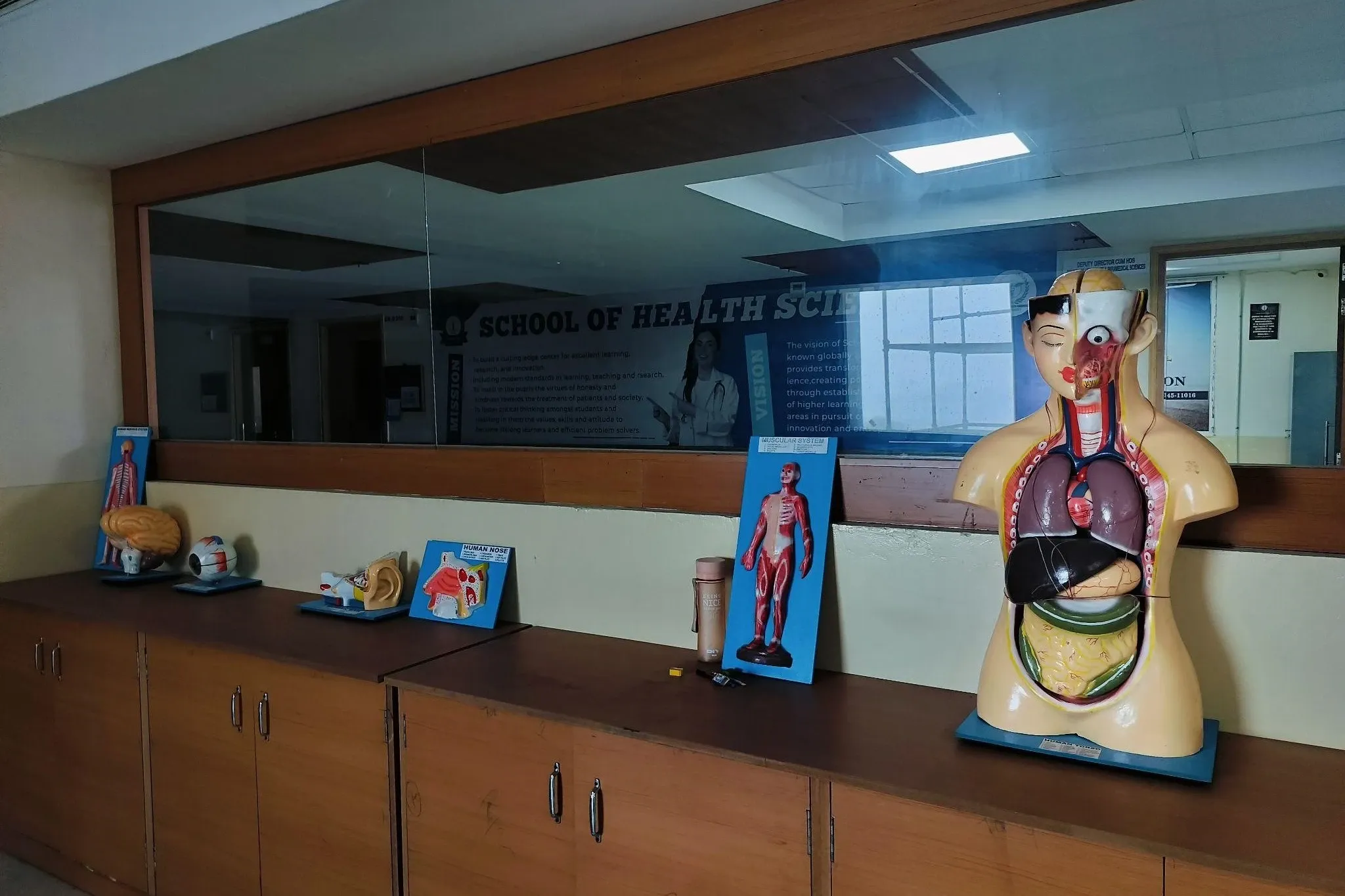
Anatomy & Physiology Lab

Anatomy & Physiology Lab

Offers anatomical models and physiological testing equipment to enhance understanding of h...

Explore
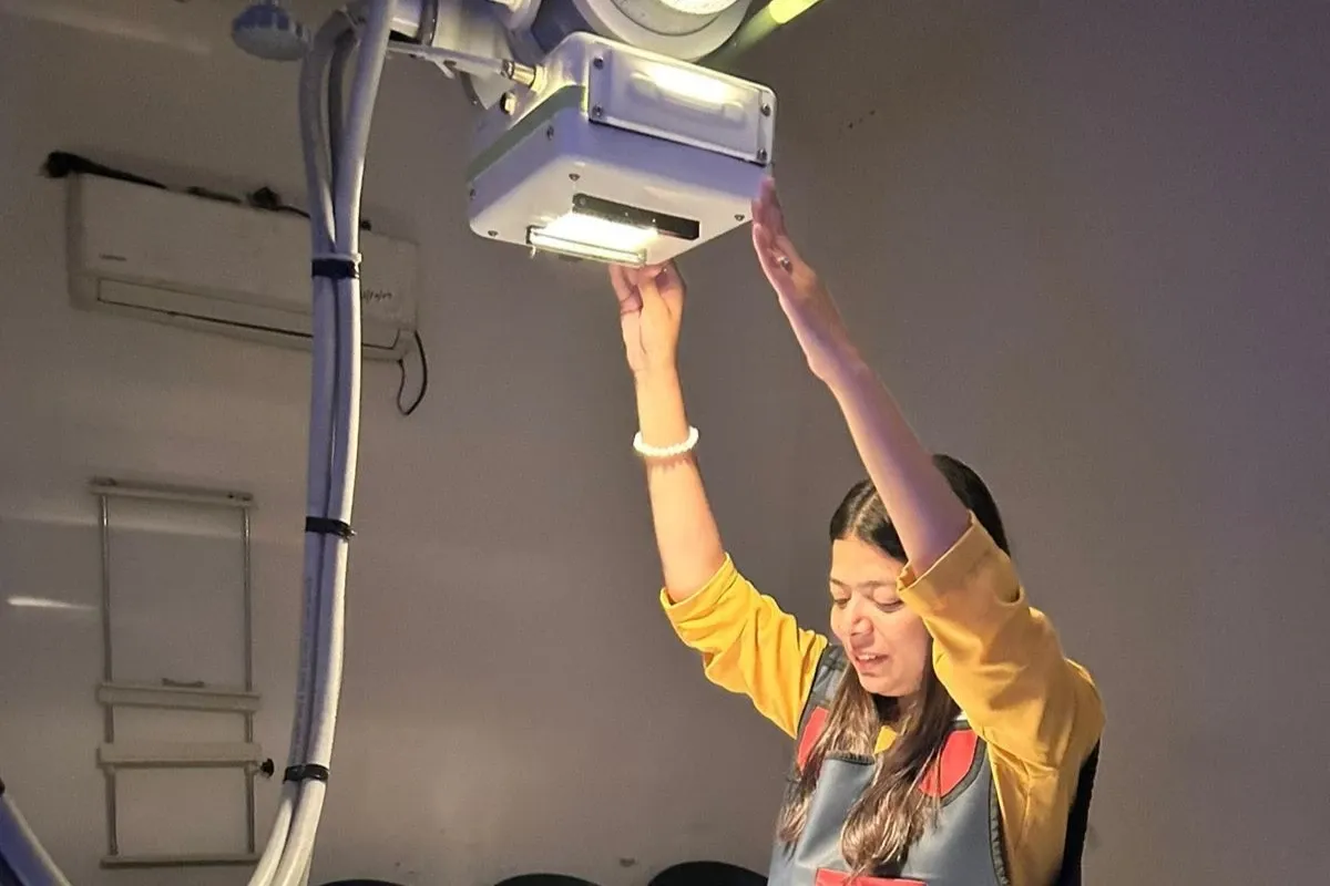
Radiology Lab

Radiology Lab

Equipped with X-ray, ultrasound, and other imaging modalities, enabling students to unders...

Explore

Backed by the Legacy of CT Group, Driven by Proven Success

20000+

Placements

800+

Collaborations

1.2Cr

Highest Placement

1800+

Recruiters

1 Lac+

Alumni

40Cr

WORTH SCHOLARSHIP

Hear From Our Students

"

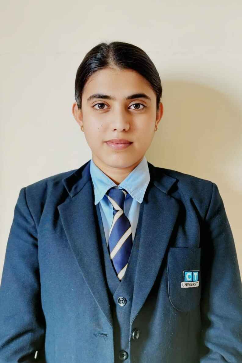
"CT University’s commitment to excellence in education is reflected in its B.Sc. Physiotherapy program. The rigorous training, focus on evidence-based practice, and emphasis on patient rehabilitation have prepared me to provide effective and ethical physiotherapy care in my career."

"

"CT University ensures a well-rounded experience by balancing academics with extracurricular activities. Various workshops, competitions, and student-led initiatives kept me engaged and motivated, helping me grow both professionally and personally."

"

"The internships and clinical training opportunities at CT University provided me with hands-on experience in hospitals and rehabilitation centers. Working under expert supervision gave me the confidence and practical skills needed to excel as a physiotherapy professional."

"

"What I appreciate most about CT University is its focus on patient-centered care and ethical practices in physiotherapy. The program not only teaches technical skills but also emphasizes compassion, safety, and responsibility in rehabilitation, shaping us into better professionals and individuals."

"

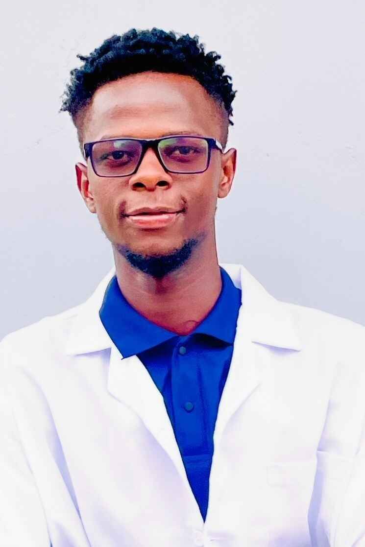
"The infrastructure at CT University is unmatched. The well-equipped labs, including digital radiography and advanced CT and MRI simulators, allowed me to practice and refine my skills. The emphasis on quality assurance and safety protocols in imaging technology ensures that graduates are well-prepared for real-world challenges."

"

"The curriculum at CT University is both comprehensive and future-focused. The B.Sc. Radiology and Imaging Technology course introduced me to advanced imaging techniques like CT, MRI, and Ultrasound, while also teaching me essential soft skills. Thanks to the guidance and placement opportunities provided, I secured a position in a reputed hospital before graduating."

"

"CT University stands out for its student-centric approach. The B.Sc. Radiology program is designed to meet industry standards, and the professors are always approachable and ready to assist. The emphasis on practical learning and internships has given me confidence and clarity in my chosen field. I highly recommend this course to anyone interested in a career in radiology."

"

"My experience at CT University has been truly transformative. The B.Sc. Radiology and Imaging Technology course provides an exceptional blend of theoretical knowledge and hands-on training. The faculty's expertise and the university's state-of-the-art labs have helped me gain a deep understanding of modern imaging techniques. This course has prepared me well for my career in medical imaging."